Brain network functional connectivity changes induced by music-induced analgesia in fibromyalgia patients
Brain network functional connectivity changes induced by music-induced analgesia in fibromyalgia patients
Mengting Pan, Jiancheng Hou, …Ying Liu Show authors
Scientific Reports volume 16, Article number: 14786 (2026) Cite this article
Abstract
Music can liberate positive power of pain management in fibromyalgia (FM) patients through multiple neural modulation. However, traditional brain research preferred to investigate the neural characteristics of music-induced analgesia (MIA) based on the seed points of functional activation, which limits the understanding of the connection states of the whole-brain functional synchronization network involved in FM’s music listening. The current study aimed to investigate the whole-brain network functional connectivity (FC) differences of resting-state functional magnetic resonance imaging (RS-fMRI) before and after music listening in FM patients using a data-driven analysis approach. Using a publicly available dataset, the RS-fMRI data from 20 FM patients were analyzed. A network-based FC approach was applied to compare intra- and inter-network FC changes across the visual network (VN), somatosensory network (SMN), ventral attention network (VAN), default mode network (DMN), and subcortical network (SC). After music listening, FM patients exhibited significant reduction in evaluation of pain intensity (PI), and also exhibited changed intra-network FC within the VAN and VN; changed inter-network FC between the VAN and DMN, between the VN and SMN or DMN, between the SMN and DMN, and between the VN and SMN or DMN, respectively. What’s more, correlations were found between post–pre changes in subjective pain ratings and post–pre changes in network FC. Positive correlations were found between PI’s reduction and the increase of inter-network FC between the right fusiform (VN) and left middle insula (SMN), and also found between the reduction of pain unpleasantness (PU) and the increase of inter-network FC between the left middle insula (SMN) and left posterior occipital cortex (DMN). The network FC results here provided new evidence to the inter/intra-networks in VN, SMN, VAN, DMN, and subcortical network, explaining that FM patients may generate cognitive processing from bottom to top and emotion regulation from top to down to realize their MIA.Download PDF
Brain network functional connectivity changes induced by music-induced analgesia in fibromyalgia patients
Mengting Pan, Jiancheng Hou, …Ying Liu Show authors
Music can liberate positive power of pain management in fibromyalgia (FM) patients through multiple neural modulation. However, traditional brain research preferred to investigate the neural characteristics of music-induced analgesia (MIA) based on the seed points of functional activation, which limits the understanding of the connection states of the whole-brain functional synchronization network involved in FM’s music listening. The current study aimed to investigate the whole-brain network functional connectivity (FC) differences of resting-state functional magnetic resonance imaging (RS-fMRI) before and after music listening in FM patients using a data-driven analysis approach. Using a publicly available dataset, the RS-fMRI data from 20 FM patients were analyzed. A network-based FC approach was applied to compare intra- and inter-network FC changes across the visual network (VN), somatosensory network (SMN), ventral attention network (VAN), default mode network (DMN), and subcortical network (SC). After music listening, FM patients exhibited significant reduction in evaluation of pain intensity (PI), and also exhibited changed intra-network FC within the VAN and VN; changed inter-network FC between the VAN and DMN, between the VN and SMN or DMN, between the SMN and DMN, and between the VN and SMN or DMN, respectively. What’s more, correlations were found between post–pre changes in subjective pain ratings and post–pre changes in network FC. Positive correlations were found between PI’s reduction and the increase of inter-network FC between the right fusiform (VN) and left middle insula (SMN), and also found between the reduction of pain unpleasantness (PU) and the increase of inter-network FC between the left middle insula (SMN) and left posterior occipital cortex (DMN). The network FC results here provided new evidence to the inter/intra-networks in VN, SMN, VAN, DMN, and subcortical network, explaining that FM patients may generate cognitive processing from bottom to top and emotion regulation from top to down to realize their MIA.
Altered resting-state functional connectome in major depressive disorder: a mega-analysis from the PsyMRI consortium
Resting-state functional connectivity predicts motor cortex stimulation-dependent pain relief in fibromyalgia syndrome patients
Functional reorganization of intranetwork and internetwork connectivity in patients with Ménière’s disease
Introduction
Fibromyalgia (FM) is present in as much as 2% to 8% of the population, is characterized by widespread pain, and is often accompanied by fatigue, memory problems and sleep disturbances1. The treatment of FM involves multiple methods, including nonpharmacological therapies (education, exercise, cognitive behavioral therapy) and pharmacological therapies (tricyclics, serotonin norepinephrine reuptake inhibitors, and gabapentinoids). Recent years, music therapy has been an important nonpharmacological method with positive biological and psychological function in pain management clinically and non-clinically2. With harmonious, emotional, and personal sound features, music can liberate therapeutic power for psychological regulation of pain (e.g., emotional regulation, motor coordination, cognitive enhancements, stress relief, etc.) and physiological intervention of pain by regulating circadian rhythm system (heart rate/blood/breathing pressure adjustment, pain control, sleep intervention, etc.), regulating hormones, such as dopamine, oxytocin and melatonin etc. Valenti3, and brain neural activities4,5.
The effect of music pain reduction, also called as music-induced analgesia (MIA), has been widely used in clinical treatment of many diseases. MIA has been demonstrated in healthy participants exposed to experimental pain6 and in patients experiencing acute7,8, postoperative9, and chronic pain10. Based on the cerebral signature for pain perception11, cognition (e.g., attentional diversion and cognitive reappraisal), emotion (e.g., affective regulation), and neurobiology (e.g., neural plasticity and functional connectivity) were proposed as the main aspects of MIA12. The aspect of cognition is often discussed in regards to MIA is distraction and attention, which can situate the analgesic capacity of music on a cognitive level13,14. Other explanations suggest that music-induced emotion is a key mechanism, which function in pleasure enhancement and anxiety reduction15,16. Among the functions of cognition, emotion, and neurobiology, the brain neural activity of music intervention to pain is a key link to explain its cognitive process and emotion regulation. The functional magnetic resonance imaging (fMRI) has demonstrated that when participants listen to their favorite music compared with no music during painful stimulation, different patterns of neural activation and functional connectivity were found in several areas of the brain, brainstem, and spinal cord, including regions within the limbic system and areas known to be involved in the descending pain-modulatory system17 and brain functional connectivity (FC) across these neural networks between regions such as the insula, thalamus, hypothalamus, amygdala and hippocampus18.
Actually, with the increasingly prominent physical and mental effects of music, more and more attention has been paid to the brain functional mechanism of MIA, which can provide important neural evidence for explaining the processing patterns and key brain regions in which music participants in pain management. By playing music to participants with their liked or disliked songs, a study found that liked music significantly rated with lowered pain ratings to acute painful stimuli compared to disliked music and no music; the lowered pain ratings were connected with brain areas encoding sensory components of pain, such as the right precentral and postcentral gyri (PreCG/PoCG), brain areas related to affective components of pain, such as the anterior cingulate cortex and bilateral putamen, and brain areas associated with motor control and avoidance reactions to pain, such as the left cerebellum19. Other studies have also found that listening to music can activate the reward circuitry20 and multiple cortical, subcortical, and cerebellar regions encompassing multiple brain networks21, which are all important neural bases for participating in MIA18.The evidence suggest that the role of music analgesia is a process involving the integration of multiple functional brain regions, so it is necessary to further explore the FC characteristics of brain22.
Pando-Naude23, examined the resting-state functional connectivity (RSFC) of MIA in FM patients. They performed RSFC seed-based correlation analyses using pain and analgesia-related regions-of-interest (ROIs) to determine the effects before and after the MIA intervention in FM and health controls (HC). The results showed that listening to music reduced pain in FM patients and this analgesic effect was related to a decrease of the RSFC on the left angular gyrus (AnG) seed with right PoCG and right precuneus, as well as a RSFC increase of the left amygdala seed with right middle frontal gyrus. The authors proposed that MIA may arise because of a top-down modulation, probably originated by distraction, relaxation, positive emotion, or a combination of these mechanisms23.
As Lunde and colleagues proposed that in-depth analysis of MIA’ mechanisms should not exist only in the knowledge but also in the use of existing analytical techniques12. The seed-based RSFC approach only provides the functional interactions between specific regions, which generally requires a defined ROI, so the results are strongly limited by ROI chosen24,25. Understanding brain activity patterns in a data-driven way can avoid limitations and flaws in assumptions, especially when the mechanism of MIA function is uncertain. Moreover, network science mainly examines the brain regions’ interactions, and it has been employed to study the human brain as one kind of the relationships of complex system. The human brain is organized to be a global network but with regular functional laws; it integrates with the large long- and the local short-range connections, in order to keep and support the high-level cognition ability26,27. As far as we known, relatively less studies have examined the effect of MIA on brain network. Therefore, the current study aimed to investigate the RS-fMRI whole-brain network FC differences by MIA in FM patients through a data-driven approach.Download PDF
Download PDF
Article
Open access
Published: 24 March 2026
Brain network functional connectivity changes induced by music-induced analgesia in fibromyalgia patients
Mengting Pan, Jiancheng Hou, …Ying Liu Show authors
Scientific Reports volume 16, Article number: 14786 (2026) Cite this article
Music can liberate positive power of pain management in fibromyalgia (FM) patients through multiple neural modulation. However, traditional brain research preferred to investigate the neural characteristics of music-induced analgesia (MIA) based on the seed points of functional activation, which limits the understanding of the connection states of the whole-brain functional synchronization network involved in FM’s music listening. The current study aimed to investigate the whole-brain network functional connectivity (FC) differences of resting-state functional magnetic resonance imaging (RS-fMRI) before and after music listening in FM patients using a data-driven analysis approach. Using a publicly available dataset, the RS-fMRI data from 20 FM patients were analyzed. A network-based FC approach was applied to compare intra- and inter-network FC changes across the visual network (VN), somatosensory network (SMN), ventral attention network (VAN), default mode network (DMN), and subcortical network (SC). After music listening, FM patients exhibited significant reduction in evaluation of pain intensity (PI), and also exhibited changed intra-network FC within the VAN and VN; changed inter-network FC between the VAN and DMN, between the VN and SMN or DMN, between the SMN and DMN, and between the VN and SMN or DMN, respectively. What’s more, correlations were found between post–pre changes in subjective pain ratings and post–pre changes in network FC. Positive correlations were found between PI’s reduction and the increase of inter-network FC between the right fusiform (VN) and left middle insula (SMN), and also found between the reduction of pain unpleasantness (PU) and the increase of inter-network FC between the left middle insula (SMN) and left posterior occipital cortex (DMN). The network FC results here provided new evidence to the inter/intra-networks in VN, SMN, VAN, DMN, and subcortical network, explaining that FM patients may generate cognitive processing from bottom to top and emotion regulation from top to down to realize their MIA.
Altered resting-state functional connectome in major depressive disorder: a mega-analysis from the PsyMRI consortium
Article Open access 07 October 2021
Resting-state functional connectivity predicts motor cortex stimulation-dependent pain relief in fibromyalgia syndrome patients
Article Open access 12 October 2022
Functional reorganization of intranetwork and internetwork connectivity in patients with Ménière’s disease
Introduction
Fibromyalgia (FM) is present in as much as 2% to 8% of the population, is characterized by widespread pain, and is often accompanied by fatigue, memory problems and sleep disturbances1. The treatment of FM involves multiple methods, including nonpharmacological therapies (education, exercise, cognitive behavioral therapy) and pharmacological therapies (tricyclics, serotonin norepinephrine reuptake inhibitors, and gabapentinoids). Recent years, music therapy has been an important nonpharmacological method with positive biological and psychological function in pain management clinically and non-clinically2. With harmonious, emotional, and personal sound features, music can liberate therapeutic power for psychological regulation of pain (e.g., emotional regulation, motor coordination, cognitive enhancements, stress relief, etc.) and physiological intervention of pain by regulating circadian rhythm system (heart rate/blood/breathing pressure adjustment, pain control, sleep intervention, etc.), regulating hormones, such as dopamine, oxytocin and melatonin etc. Valenti3, and brain neural activities4,5.
The effect of music pain reduction, also called as music-induced analgesia (MIA), has been widely used in clinical treatment of many diseases. MIA has been demonstrated in healthy participants exposed to experimental pain6 and in patients experiencing acute7,8, postoperative9, and chronic pain10. Based on the cerebral signature for pain perception11, cognition (e.g., attentional diversion and cognitive reappraisal), emotion (e.g., affective regulation), and neurobiology (e.g., neural plasticity and functional connectivity) were proposed as the main aspects of MIA12. The aspect of cognition is often discussed in regards to MIA is distraction and attention, which can situate the analgesic capacity of music on a cognitive level13,14. Other explanations suggest that music-induced emotion is a key mechanism, which function in pleasure enhancement and anxiety reduction15,16. Among the functions of cognition, emotion, and neurobiology, the brain neural activity of music intervention to pain is a key link to explain its cognitive process and emotion regulation. The functional magnetic resonance imaging (fMRI) has demonstrated that when participants listen to their favorite music compared with no music during painful stimulation, different patterns of neural activation and functional connectivity were found in several areas of the brain, brainstem, and spinal cord, including regions within the limbic system and areas known to be involved in the descending pain-modulatory system17 and brain functional connectivity (FC) across these neural networks between regions such as the insula, thalamus, hypothalamus, amygdala and hippocampus18.
Actually, with the increasingly prominent physical and mental effects of music, more and more attention has been paid to the brain functional mechanism of MIA, which can provide important neural evidence for explaining the processing patterns and key brain regions in which music participants in pain management. By playing music to participants with their liked or disliked songs, a study found that liked music significantly rated with lowered pain ratings to acute painful stimuli compared to disliked music and no music; the lowered pain ratings were connected with brain areas encoding sensory components of pain, such as the right precentral and postcentral gyri (PreCG/PoCG), brain areas related to affective components of pain, such as the anterior cingulate cortex and bilateral putamen, and brain areas associated with motor control and avoidance reactions to pain, such as the left cerebellum19. Other studies have also found that listening to music can activate the reward circuitry20 and multiple cortical, subcortical, and cerebellar regions encompassing multiple brain networks21, which are all important neural bases for participating in MIA18.The evidence suggest that the role of music analgesia is a process involving the integration of multiple functional brain regions, so it is necessary to further explore the FC characteristics of brain22.
Pando-Naude23, examined the resting-state functional connectivity (RSFC) of MIA in FM patients. They performed RSFC seed-based correlation analyses using pain and analgesia-related regions-of-interest (ROIs) to determine the effects before and after the MIA intervention in FM and health controls (HC). The results showed that listening to music reduced pain in FM patients and this analgesic effect was related to a decrease of the RSFC on the left angular gyrus (AnG) seed with right PoCG and right precuneus, as well as a RSFC increase of the left amygdala seed with right middle frontal gyrus. The authors proposed that MIA may arise because of a top-down modulation, probably originated by distraction, relaxation, positive emotion, or a combination of these mechanisms23.
As Lunde and colleagues proposed that in-depth analysis of MIA’ mechanisms should not exist only in the knowledge but also in the use of existing analytical techniques12. The seed-based RSFC approach only provides the functional interactions between specific regions, which generally requires a defined ROI, so the results are strongly limited by ROI chosen24,25. Understanding brain activity patterns in a data-driven way can avoid limitations and flaws in assumptions, especially when the mechanism of MIA function is uncertain. Moreover, network science mainly examines the brain regions’ interactions, and it has been employed to study the human brain as one kind of the relationships of complex system. The human brain is organized to be a global network but with regular functional laws; it integrates with the large long- and the local short-range connections, in order to keep and support the high-level cognition ability26,27. As far as we known, relatively less studies have examined the effect of MIA on brain network. Therefore, the current study aimed to investigate the RS-fMRI whole-brain network FC differences by MIA in FM patients through a data-driven approach.
The participants’ information, and their RS-fMRI and anatomical T1 dataset, were obtained from a public dataset via OpenNeuro with accession number There were twenty female FM patients (mean age = 46.4 ± 12.4 years old, ranged from 22 to 70 years). Ethical approval was granted by the Bioethics Committee of the Instituto de Neurobiología, UNAM. The inclusion and exclusion criteria followed those outlined by Pando-Naude et al.23,.
Stimulus materials
Prior to the study, participants provided a list of songs or artists that they would like to listen during the experiment. The Songs were familiar, very pleasant, and slow. The slow pace was defined as a tempo of < 120 beats per minute (bpm), determined by the experimenters using a metronome. Participants reported how pleasant the song was on a 11-point verbal scale (0 = unpleasant, 10 = very pleasant), and to be selected, the song had to be rated at least 9–10. When provided with only the artist’s name, the experimenter selected the songs based on two fixed acoustic criteria: harmony (pleasurable), participants’ oral reporting, and slow tempo. Based on a previous study, music-induced analgesia for chronic pain was higher when participants chose familiar and happy music, making self-selection and familiarity an important mechanism for this effect23.
Design and paradigm
Auditory stimuli were used the NordicNeuroLab AS (Bergen, Norway) MRI safety headphones, and their presenting orders were balanced among the participants in order to avoid sequential effects. Each participant underwent two RS-fMRI acquisitions that were performed before (pre-) and after (post-) music listening, each lasting five minutes. Inside the MRI scanner, participants listened to a 5-minute segment of familiar, slow paced, and highly pleasant music, and no imaging was recorded during music listening. Before the RS-fMRI acquisition, the anatomical T1-weighted data were acquired23.
Meantime, two behavioral tasks, the pain intensity (PI) and the pain unpleasantness (PU), were tested while the FM patients were in the MRI scanner and using the verbal rating scale (0 = no pain, 10 = worst pain possible). PI measures the pain sensory, and the PU measures the evaluative and emotional dimension of pain23. The two tasks were performed before and after the music stimuli.
MRI data acquisition
The original neuroimaging dataset was acquired on a 3.0 Tesla GE Discovery MR750 scanner (HD, General Electric Healthcare, Waukesha, WI, USA) and a commercial 32-channel head coil array. The parameters for RS-fMRI data were: TR = 3000 ms, TE = 40 ms, flip angle = 90°, field of view = 256 × 256 mm2, voxel size = 2 × 2 mm, number of slices = 43, matrix = 128 × 128, slice thickness = 3 mm, gap = 0 mm. The parameters for anatomical T1-weighted data were: TR = 7.7 ms, TE = 3.2 ms, flip angle = 12°, field of view = 256 × 256 mm2, matrix = 256 × 256, slice thickness = 1 mm, number of slices = 168, gap = 0 mm.
Data preprocessing
Preprocessing for RS-fMRI data were performed using the Data Processing and Analysis of Brain Imaging (DPABI) toolbox version 6.0 (http://rfmri.org/dpabi). This toolbox involves one main function called Data Processing Assistant for Resting-state fMRI Advanced Edition toolbox (DPARSF V5.3)28,29, which is a convenient plug-in software that works with Matlab and Statistical Parametric Mapping (SPM, version 12) (https://www.fil.ion.ucl.ac.uk/spm/software/spm12/). The original data was firstly arranged; the first five volumes were ignored so that participants could get used to scanner noise. The preprocessing steps, in order, includes slice timing, realignment, regressing out head motion parameters (scrubbing with Friston 24-parameter model regression) and normalization (spatial normalization to the MNI template, resampling voxel size of 3 × 3 × 3 mm)30,31. The symmetric correlation matrix (considered as spontaneous neural connectivity) for a 142 × 141 network, which include 142 regions/nodes with the Dosenbach atlas across whole brain32, was generated in each participant. Therefore, each participant had 20,022 unique pairwise RSFC in total, but only half of the pairwise RSFC (10,011) within the network was used for next network construction, because the top right half and bottom left half were the same in matrix.
Network functional connectivity construction
Each participant’s network functional connectivity (FC) was constructed with the DPABINet V1.1 that is included in DPABI V6.0. The 142 nodes in the Dosenbach atlas were classified into seven subnetworks: visual network (VN), somatosensory network (SMN), ventral attention network (VAN), dorsal attention network (DAN), default mode network (DMN), subcortical network (SC) and frontoparietal network (FPN)33. Based on the 142 × 141 matrix generated at the preprocessing step, the network FC for any pair of two nodes was calculated as Pearson’s linear correlation coefficient, and the Fisher-z transformation was then used for the symmetric correlation matrix. It needs to be mentioned that the smoothing was not used to the brain network analysis because the core objective of brain network analysis is to accurately measure the functional connectivity relationships among discrete brain regions34; if smoothing is used in brain network analysis, it will have serious problems such as that the signals between brain regions are no longer pure and will artificially and falsely increase the functional connection value between two brain nodes34,35; or that smoothing can blur the spatial boundaries of the networks, reducing the specificity of internal connections within the network and the clarity of separation between networks; or that smoothing can make brain nodes no longer independent at the signal level34,36.
Statistical analysis
The statistical analysis was performed in DPABINet. The paired t-test (after vs. before music listening) was conducted to compare the network FC differences between the post- vs. pre-music listening in FM patients. The gender and age were considered as covariates. The multiple comparison correction was used the permutation false discovery rate (FDR) corrected p <.05 in DPABINet, and the results were visualized by DPABINet Viewer. Moreover, the correlation analysis based on Pearson’s correlation between the PI (or the PU; the difference value between post- minus pre-music listening) and the network FC (the difference value between post- minus pre-music listening) was set to p <.05 with SPSS software version 23 without multiple-comparison corrections.
Results
Behavioral test
The paired t-test showed that after music listening, the FM patients’ PI was significantly decreased, and the PU was marginally decreased compared to before music listening.
Network functional connectivity changes
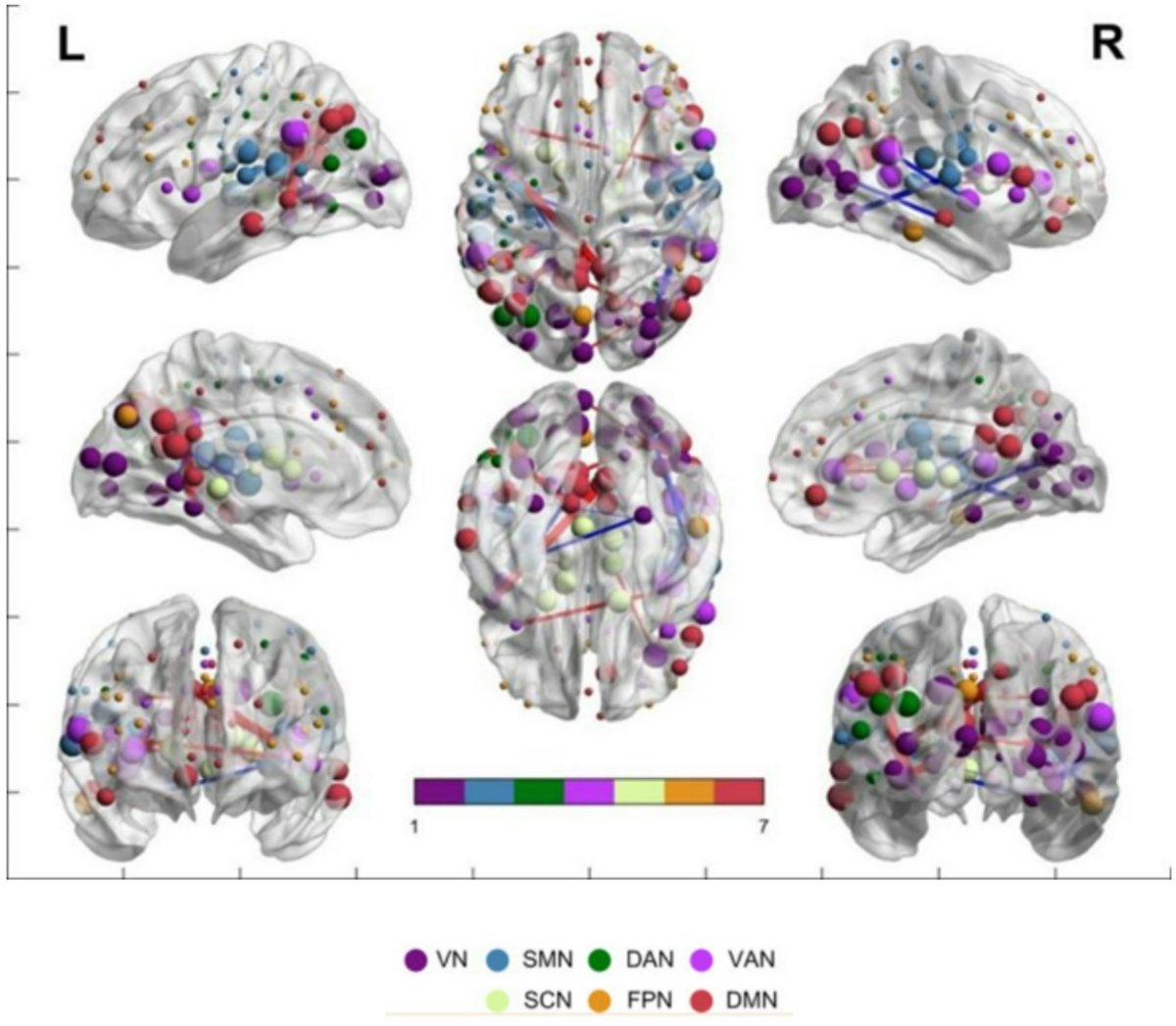
Table 2; Figs. 1 and 2 show the significant intra- and inter-network FC differences between post- vs. pre-music listening in FM patients. Compared to pre-music listening, post-music listening induced enhanced intra-network FC between the right ventral frontal cortex and right ventromedial prefrontal cortex, or the left anterior insula within the VAN; between the left posterior occipital cortex and right temporal cortex within the VN; but induced decreased intra-network FC between the right parietal lobule and right middle insula within the VAN.
Compared to pre-music listening, post-music listening induced enhanced inter-network FC between the VAN and DMN; between the VN and SMN or DMN, respectively. Moreover, compared to pre-music listening, post-music listening induced decreased inter-network FC between the SMN and DMN; between the VN and SMN or DMN, respectively.References
Clauw, D. J. M. Fibromyalgia: A clinical review. JAMA 311(15), 1547–1555. https://doi.org/10.1001/jama.2014.3266 (2014).
Finn, S. & Fancourt, D. The biological impact of listening to music in clinical and nonclinical settings: A systematic review. Prog Brain Res. 237, 173–200. https://doi.org/10.1016/bs.pbr.2018.03.007 (2018)
Valenti, V. E. et al. Auditory stimulation and cardiac autonomic regulation. Clinics (Sao Paulo, Brazil) https://doi.org/10.6061/clinics/2012(08)16 (2012).